Discover the complete Eye Anatomy 2026 guide. Learn about the cornea, retina, iris, lens, optic nerve, eye functions, common disorders, and the latest advances in eye health.
Eye anatomy 2026
Introduction
The human eye is one of the most complex and fascinating organs in the body. Often compared to a high-resolution camera, the eye captures light, processes visual information, and sends signals to the brain, allowing us to see the world around us. Understanding eye anatomy is essential for students, healthcare professionals, and anyone interested in maintaining healthy vision.
What Eye Anatomy?
Eye anatomy refers to the structure and organization of the eye and its associated tissues. Each part of the eye performs a specialized function that contributes to vision. Together, these structures collect light, focus images, convert light into electrical signals, and transmit those signals to the brain.
External Anatomy of the Eye
1. Eyelids
The eyelids protect the eyes from dust, debris, and excessive light. Blinking helps spread tears across the eye surface, keeping it moist and clean.
Functions:
Protect the eye
Spread tears evenly
Remove foreign particles
Prevent dryness
2. Eyelashes
Eyelashes act as a protective barrier that prevents dust and small particles from entering the eye.
Functions:
Detect foreign objects
Trigger blink reflex
Reduce airborne debris exposure
3. Eyebrows
Eyebrows help divert sweat and rain away from the eyes while also contributing to facial expressions.
Internal Anatomy of the Eye
1. Cornea
The cornea is the transparent front layer of the eye. It serves as the primary focusing structure and contributes significantly to the eye’s refractive power.
Functions:
Focus incoming light
Protect internal eye structures
Filter some ultraviolet rays
Cornea in Eye Anatomy: Structure, Functions, and Importance
What is the Cornea?
The cornea is the transparent, dome-shaped front surface of the eye that covers the iris, pupil, and anterior chamber. It is one of the most important parts of eye anatomy because it helps focus light entering the eye, allowing us to see clearly.
Location of the Cornea
The cornea is located at the very front of the eye. It acts like a clear window through which light enters before passing through the pupil and lens.
Light Path Through the Eye
Cornea
Aqueous Humor
Pupil
Lens
Vitreous Humor
Retina
Optic Nerve
Main Functions of the Cornea
1. Focuses Light
The cornea provides approximately 65–75% of the eye’s focusing power. It bends (refracts) incoming light so that it reaches the retina correctly.
2. Protects the Eye
The cornea serves as a protective barrier against:
Dust
Germs
Dirt
Harmful UV rays
Small foreign objects
3. Maintains Clear Vision
A healthy, transparent cornea is essential for sharp and clear vision. Any damage or clouding can significantly affect eyesight.
A scratch on the cornea that causes pain, redness, and sensitivity to light.
2. Keratitis
Inflammation of the cornea, often caused by infection.
3. Keratoconus
A condition where the cornea becomes thin and bulges outward into a cone shape.
4. Corneal Ulcer
An open sore on the cornea that can threaten vision if untreated.
5. Corneal Dystrophy
Inherited disorders affecting corneal clarity and function.
Symptoms of Corneal Problems
Blurred vision
Eye pain
Redness
Excessive tearing
Light sensitivity
Feeling that something is in the eye
How to Keep Your Cornea Healthy
Wear UV-protective sunglasses.
Use contact lenses properly.
Avoid rubbing your eyes excessively.
Eat foods rich in Vitamin A and Omega-3.
Get regular eye examinations.
Use protective eyewear during sports and hazardous work.
Interesting Facts About the Cornea
The cornea contains no blood vessels.
It receives oxygen directly from the air and tears.
It is one of the most sensitive tissues in the human body.
The cornea can heal quickly from minor injuries.
Conclusion
The cornea is the clear front layer of the eye that plays a crucial role in focusing light and protecting the eye from external damage. Maintaining a healthy cornea is essential for clear vision and overall eye health. Understanding the cornea’s structure and function helps us appreciate its importance in the complex system of human eye anatomy.
2. Sclera
The sclera is the white outer covering of the eyeball.
Functions:
Maintains eye shape
Provides structural support
Protects internal tissues
3. Conjunctiva
The conjunctiva is a thin membrane covering the sclera and inner eyelids.
Functions:
Lubricates the eye
Protects against infection
Supports immune defense
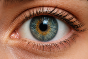
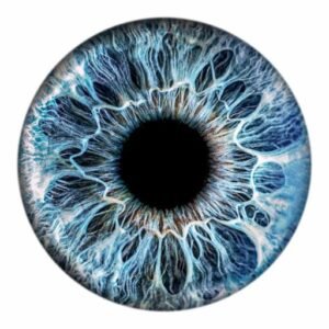
4. Iris
The iris is the colored part of the eye.
Functions:
Controls pupil size
Regulates light entry
Determines eye color
5. Pupil
The pupil is the black opening in the center of the iris.
Functions:
Allows light into the eye
Adjusts to lighting conditions
Lens Anatomy
Crystalline Lens
The lens is a transparent structure located behind the iris.
Functions:
Focuses light onto the retina
Adjusts focus for near and distant objects
Changes shape during accommodation
Common Lens Disorders:
Cataracts
Presbyopia
Lens dislocation
Retina: The Vision Processing Center
The retina is a light-sensitive layer located at the back of the eye
Layers of the Retina
Rod Cells
Responsible for:
Night vision
Peripheral vision
Motion detection
Cone Cells
Responsible for:
Color vision
Detailed vision
Daylight vision
Macula
The macula is the central area of the retina responsible for detailed vision.
Fovea
The fovea provides the sharpest vision and contains the highest concentration of cone cells.
Optic Nerve
The optic nerve carries visual signals from the retina to the brain.
Functions:
Transmits visual information
Connects eye and brain
Enables image interpretation
Disorders:
Glaucoma
Optic neuritis
Optic nerve atrophy
Vitreous Humor
The vitreous humor is a gel-like substance filling the back chamber of the eye.
Functions:
Maintains eye shape
Supports the retina
Allows light transmission
Aqueous Humor
Aqueous humor is a clear fluid located in the front part of the eye.
Functions:
Nourishes the cornea and lens
Maintains eye pressure
Removes metabolic waste
Eye Muscles and Movement
Six extraocular muscles control eye movement.
Superior Rectus
Moves the eye upward.
Inferior Rectus
Moves the eye downward.
Medial Rectus
Moves the eye inward.
Lateral Rectus
Moves the eye outward.
Superior Oblique
Assists with rotational movement.
Inferior Oblique
Supports upward and rotational movement.
How Vision Works
The visual process occurs in several steps:
Light enters through the cornea.
The pupil regulates incoming light.
The lens focuses light.
Light reaches the retina.
Photoreceptors convert light into electrical signals.
Signals travel through the optic nerve.
The brain interprets the image.
Blood Supply of the Eye
The eye receives oxygen and nutrients from branches of the ophthalmic artery.
Key Vessels:
Central retinal artery
Ciliary arteries
Choroidal circulation
Proper blood flow is essential for retinal health and vision preservation.
Common Eye Diseases
Cataracts
A clouding of the lens causing blurry vision.
Symptoms:
Blurred vision
Light sensitivity
Difficulty seeing at night
Glaucoma
Damage to the optic nerve, often associated with increased eye pressure.
Symptoms:
Peripheral vision loss
Eye pain
Vision impairment
Age-Related Macular Degeneration (AMD)
Affects the macula and central vision.
Symptoms:
Distorted vision
Difficulty reading
Central blind spots
Diabetic Retinopathy
A complication of diabetes affecting retinal blood vessels.
Symptoms:
Floaters
Blurred vision
Vision loss
Latest Eye Anatomy and Vision Advances in 2026
Recent developments in eye care include:
AI-powered retinal imaging
Advanced cataract surgery techniques
Gene therapy for inherited retinal diseases
Smart contact lenses
Improved glaucoma monitoring systems
Enhanced OCT (Optical Coherence Tomography) imaging
These innovations continue to improve diagnosis, treatment, and prevention of vision-related disorders.
Eye Health Tips
Maintain Healthy Vision
Eat leafy green vegetables
Consume omega-3-rich foods
Wear UV-protective sunglasses
Take regular screen breaks
Stay hydrated
Schedule routine eye examinations
Control blood sugar levels
Avoid smoking
How to Maintain Healthy Eyes
Maintaining good eye health is important for clear vision and preventing eye diseases. Here are some effective tips:
1. Eat Eye-Friendly Foods
Include foods rich in:
Vitamin A: Carrots, sweet potatoes, spinach
Vitamin C: Oranges, lemons, guava
Vitamin E: Almonds, sunflower seeds
Omega-3 Fatty Acids: Fish, flaxseeds, walnuts
Lutein and Zeaxanthin: Kale, spinach, broccoli
2. Stay Hydrated
Drink plenty of water throughout the day to prevent dry eyes and maintain proper tear production.
3. Follow the 20-20-20 Rule
If you use a computer or mobile device frequently:
Every 20 minutes
Look at something 20 feet away
For at least 20 seconds
This helps reduce digital eye strain.
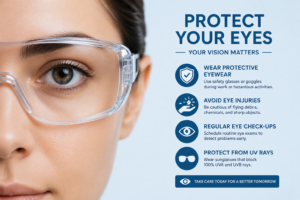
4. Protect Your Eyes from UV Rays
Wear sunglasses that block 100% UVA and UVB rays when outdoors.
5. Get Enough Sleep
Adults should aim for 7–9 hours of sleep per night. Lack of sleep can cause eye fatigue, dryness, and irritation.
6. Avoid Smoking
Smoking increases the risk of:
Cataracts
Macular degeneration
Optic nerve damage
7. Practice Good Eye Hygiene
Wash your hands before touching your eyes.
Remove makeup before sleeping.
Clean contact lenses properly.
8. Reduce Screen Brightness
Adjust screen brightness and use proper lighting while working to reduce eye strain.
9. Exercise Regularly
Physical activity improves blood circulation, which helps deliver oxygen and nutrients to the eyes.
10. Get Regular Eye Exams
Visit an eye specialist at least once every 1–2 years, even if your vision seems normal.
Warning Signs to Watch For
Seek medical attention if you experience:
Sudden vision loss
Persistent eye pain
Flashes of light
Double vision
Severe redness or swelling
Protecting your eyes is important for maintaining good vision and preventing eye diseases. Here are some practical tips:
1. Follow the 20-20-20 Rule
If you spend a lot of time looking at screens:
Every 20 minutes
Look at something 20 feet away
For at least 20 seconds
This helps reduce digital eye strain.
2. Eat Eye-Healthy Foods
Include foods rich in:
Vitamin A: carrots, sweet potatoes, spinach
Vitamin C: oranges, strawberries
Vitamin E: almonds, sunflower seeds
Omega-3 fatty acids: fish, flaxseeds
Lutein and zeaxanthin: kale, spinach, broccoli
3. Wear UV-Protective Sunglasses
Choose sunglasses that block:
99%–100% of UVA rays
99%–100% of UVB rays
This helps protect against cataracts and other eye damage.
4. Keep Your Eyes Moist
Blink regularly when using digital devices.
Use artificial tears if your eyes feel dry.
Stay hydrated by drinking enough water.
5. Reduce Screen Strain
Adjust screen brightness to match your surroundings.
Keep screens about an arm’s length away.
Increase text size if needed.
Use blue-light filters during evening hours.
6. Get Enough Sleep
Adults should aim for 7–9 hours of sleep each night. Proper rest helps your eyes recover from daily strain.
7. Avoid Smoking
Smoking increases the risk of:
Cataracts
Dry eyes
Age-related macular degeneration
8. Practice Good Eye Hygiene
Wash your hands before touching your eyes.
Remove eye makeup before sleeping.
Clean contact lenses properly if you wear them.
9. Exercise Regularly
Physical activity improves blood circulation, which helps deliver oxygen and nutrients to your eyes.
10. Get Regular Eye Exams
Visit an eye doctor every 1–2 years, or more often if you have:
Diabetes
High blood pressure
A family history of eye disease
Warning Signs That Need Immediate Medical Attention
Sudden vision loss
Eye pain
Flashes of light
Double vision
Severe redness or swelling
Early treatment can prevent permanent vision damage.
Conclusion
Eye anatomy is a remarkable combination of specialized structures working together to create vision. From the transparent cornea to the highly sensitive retina and optic nerve, each component plays a vital role in visual perception. Understanding the anatomy of the eye helps improve awareness of eye health, supports early disease detection, and encourages proper vision care. As technology advances in 2026, new diagnostic and treatment methods continue to enhance our ability to protect and preserve sight for future generations.
SEO Keywords
Eye Anatomy, Human Eye Anatomy, Eye Structure, Retina Anatomy, Cornea Function, Optic Nerve, Iris Anatomy, Lens of the Eye, Eye Health 2026, Vision System, Eye Parts and Functions, Retina Structure, Human Vision, Eye Diseases, Eye Care Guide
Tags
Eye Anatomy, Human Eye, Retina, Cornea, Iris, Pupil, Lens, Optic Nerve, Vision, Eye Health, Eye Care, Medical Education, Ophthalmology, Anatomy, Human Body, Eye Structure, Retina Function, Vision Science, Health Guide, Medical Science